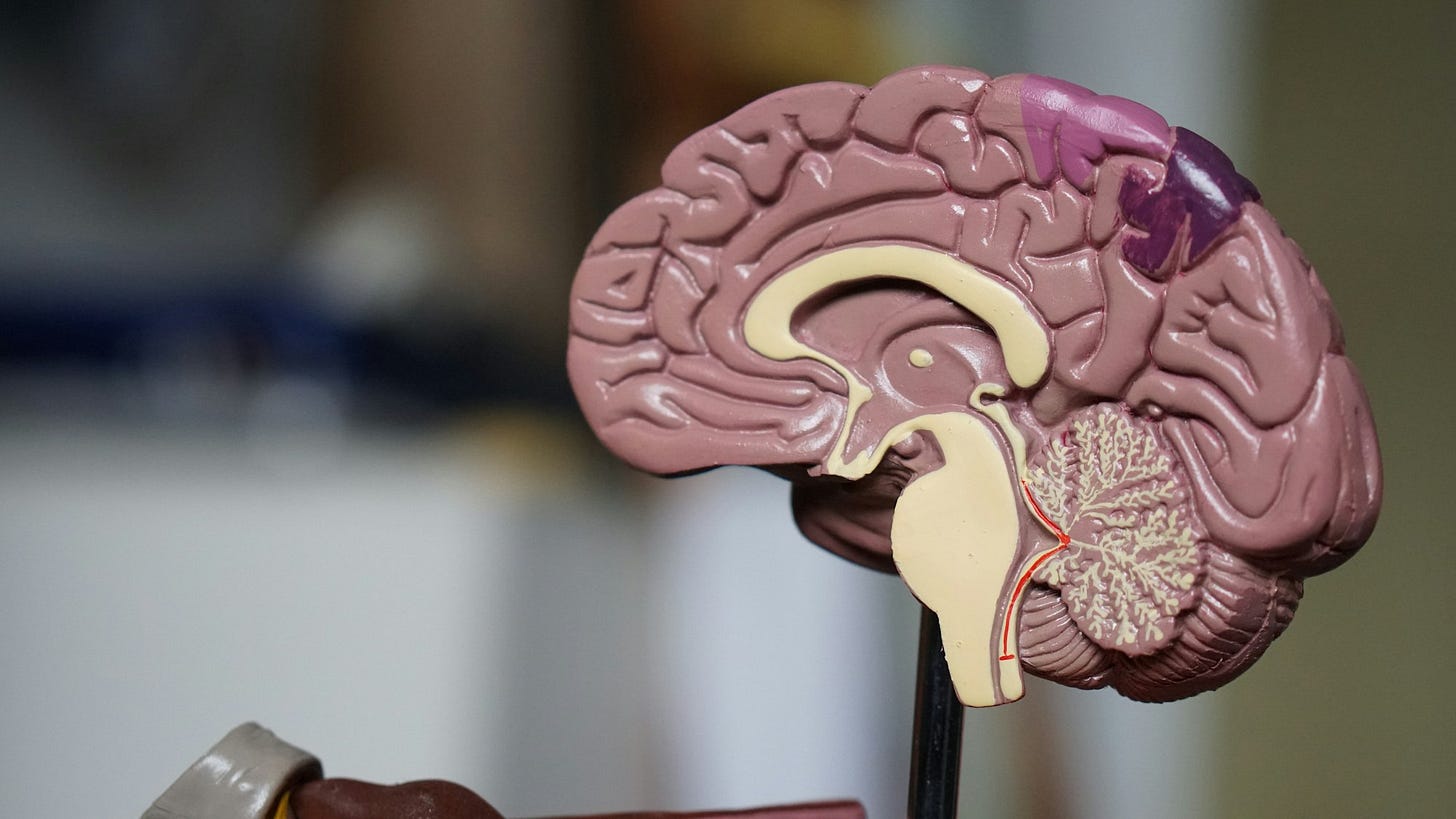
Ninety percent of your brain is (not) useless
A close look at the idea that most of the brain is superfluous space, with a review of people who get by with extraordinarily small brain mass.

I often mention the "Sherlock Holmes theory of mind" in my classes. The idea is that the mind has a limited (and small) capacity, so that filling it with useless information takes away space that could be better devoted to useful knowledge. The opposite number to the "Sherlock Holmes theory", naturally, is the "Einstein theory of mind"—namely, the idea that the ordinary human uses only 10 percent of his or her brain.
At the outset, I should point out that the Einstein story is a total urban myth, which lately has been used to great effect by psychics, self-actualization seminar leaders, and various other charlatans. There is no record that Einstein ever wrote or said anything about the useful percentage of his or anyone else's brain.
Neuroscientist and skeptic Barry Beyerstein (1999) traced the myth ultimately to the "New Thought" movement, which "blossomed following the U.S. Civil War among the prosperity-obsessed yet anxiety-ridden middle classes" (Beyerstein 1999:6, paraphrasing Meyer 1965). Popularizers of the idea ranged from devotees of numerology to self-help guru Dale Carnegie, who himself cited William James for the idea. Beyerstein himself investigated whether there is any truth to the Einstein story (there isn't), and with some assistance was able to track down the idea in the text of William James' public lectures.
But despite the fact that the myth itself is bunk, various neurologists have from time to time advanced evidence to support this myth. I'm writing a bit about this, because I've been thinking about the context of the LB1 brain and the hypothesis that it might have had "advanced" capacities. It is obvious that natural selection could reduce the size of the human brain by half or more with functional loss—this would simply be a reversal of the Pleistocene evolution of brain size. The basic question is whether natural selection could reduce the brain size of a hominid population to half or less, without reducing some cognitive capabilities. Is it possible to build a leaner, meaner brain?
Should we have a strong opinion about this? So much about the brain is unknown, that the hypothesis may simply be untestable. How could we demonstrate that a population with hobbit-sized brains could not have been just as cognitively adept as some modern human group? It is a daunting question to try to answer. I'm not going to try to answer it here.
What I do want to do is give an account of some of the examples from the neuroscience literature that people have used to support the idea that brains could evolve to be smaller without functional compromise. Many distinct conditions lead to small brain tissue volume, including hydrocephalus, microcephaly, and deliberate hemispherectomy. A number of studies have claimed that people with profound reductions in brain volume—down to as little as 150ml—nevertheless have entirely normal cognitive function. This would be a real-world manifestation of the 10 percent myth: a person using literally 10 percent of the average brain volume to live an ordinary life.
But the research in this area is essentially anecdote leavened by CT and psychometric results that might—or might not—show what they are proposed to demonstrate. I'm going to focus here on two examples, the work of John Lorber on profound reduction in brain volume associated with hydrocephalus, and hemispherectomy. There are several others that deserve some treatment, including clinical microcephaly presenting with normal intelligence, although in many such cases we are merely looking at unusually small brain volumes and not reductions to half or more of the average size. The examples I'm examining here are some of the most extreme claims of cognitive performance with minimal brain size.
I should mention some caveats at the outset. Most cases of pathology are not as extreme in their effects on brain size as the exceptional cases that have sometimes been cited. For example, the majority of people diagnosed with hydrocephalus actually have only a small reduction in brain volume as a result of slight increase in size of the fluid-filled ventricles. Hydrocephalus is an eminently treatable condition, and many patients have no cognitive deficits at all—even those for whom the proper treatment is to do nothing. Moreover, there is no debate among neurologists that the mind can sometimes recover, heal, or form around devastating anatomical challenges. The cases reviewed here are extreme ones, where brain volume is reduced to a fraction of its usual size.
Also, it is hard to avoid using the term "normal" in the context of pathology, but we should recognize that "normal" includes a substantial degree of variability. There is no single concept of normalcy applicable to human cognition. The best we can do is apply psychometric and performance measures to samples of people to assess the characteristics of human variability. Hence, when someone asserts that an individual has "normal" cognitive performance, it is far from obvious what they mean. Does it mean that they fit within 2 standard deviations of the mean? Does it mean "average"? With these conditions that often manifest during early development, "normal" often means within some specified distance of the average development expected for a given age. But someone with considerable cognitive deficits may nevertheless be termed "normal" in respect of some developmental scale—for instance, "normal" when accounting for a 2-year developmental delay.
So, we must approach the idea of "normal" cognition with some skepticism. From an evolutionary point of view, the only "normal" that matters is respect to fitness within a population. And there remains no well-defined connection between cognition and fitness at all, beyond the evidence of our current cognitive adaptations as the result of past selection. This is another area in which our hypothesis is ill-defined, making it very difficult to test.
John Lorber and hydrocephalus
The most well-known neurologist who argued that brain size could radically shrink without functional compromise was John Lorber. Lorber's work with patients of hydrocephalus received substantial public attention, including a documentary film and a profile by writer Roger Lewin in Science. Lorber studied hundreds of cases of hydrocephaly, but the value—or lack of value—of his evidence is illustrated by a single anecdote:
"There's a young student at this university," says Lorber, "who has an IQ of 126, has gained a first-class honors degree in mathematics, and is socially completely normal. And yet the boy has virtually no brain." The student's physician at the university noticed that the youth had a slightly larger than normal head, and so referred him to Lorber, simply out of interest. "When we did a brain scan on him," Lorber recalls, "we saw that instead of the normal 4.5-centimeter thickness of brain tissue between the ventricles and the cortical surface, there was just a thin layer of mantle measuring a millimeter or so. His cranium is filled mainly with cerebrospinal fluid" (Lewin 1980:1232).
Much of the apparent "surprise" in this case owes to the presumably small total volume of the brain, and the cerebrum in particular. Lorber interpreted the small cortical volume coupled with normal—or even "superior"— cognitive performance as especially surprising.
Let's take this claim part by part. What exactly is surprising about it?
1. The allegedly small cortical thickness: Medical CT scans, particularly those taken during the 1970's, do not have the resolution sufficient to accurately measure tissue thickness down to submillimeter values. A millimeter is about the limit of the resolution. A phrase like "measuring a millimeter or so" should therefore be taken generously: the cortex is very thin, possibly at the limits of detection for the equipment.
Even so, no CT or MRI images I have seen of hydrocephalics—an admittedly small number—have shown a cortex that is uniformly thin. The enlarged ventricles reduce the cortical volume by hydrostatic pressure, but this pressure is not uniform around the entire cortex circumference. A single thin layer of tissue would require an extraordinary mechanism to attain. In this and some other of Lorber's patients, the hydrocephaly apparently had an onset later than suture closure, so that the enlarged ventricles did not have a major effect on cranial circumference. This itself is an unusual etiology, but consider what this story omits. What about the size of noncortical brain structures, including the cerebellum? What about the distribution of thickness of the cortex? Are there regions that are thinner than others? It is hard to interpret the "millimeter or so".
The cerebral cortex of a "normal" individual only averages 2mm thick. With a total surface area of around 880cm2, this leads to a total cortical volume of only 191ml, again for a normal individual. The impressive thinness of the cortex comes from its internal structure, organized in humans as in most mammals with only 6 layers of neurons. Hydrocephalus mainly decreases the volume of the white matter, composed of myelinated axons connecting brain regions to each other. White matter damage is the main cause of long-term cognitive deficits resulting from hydrocephalus (discussed a bit more below), but a substantial degree of white matter damage is possible in many brain pathologies (including Alzheimer's) with cognitive impairment that is either limited in extent or limited to a few functions.
2. High cognitive performance with low cortical volume:
Consider this passage from Lewin's (1980) review:
Lorber divides the patients into four categories: those with minimally enlarged ventricles; those whose ventricles fill 50 to 70 percent of the cranium; those in which the ventricles fill between 70 and 90 percent of the intracranial space; and the most severe group, in which ventricle expansion fills 95 percent of the cranium. Many of the individuals in this last group, which forms just less than 10 percent of the total sample, are severely disabled, but half of them have IQ's greater than 100. This group provides some of the most dramatic examples of apparently normal function against all odds (Lewin 1980:1232).
No one can dispute that the brain is capable of amazing feats of repair and reorganization, which sometimes permit normal function in the face of profound pathology. But the notion that "half" of the patients where ventricle expansion is greater than 95 percent of the cranium have IQ's greater than 100 is mathematically implausible. The definition of IQ is that the mean is 100. This means that only half of people withoutventricle expansion have IQ over 100. Lorber seems to have claimed that the most severe cases of hydrocephalus actually see an increase in the proportion of high-IQ individuals, despite "many" being severely disabled. I'm not saying it's impossible, but like "a millimeter or so," this is the kind of statistic that deserves skepticism.
3. "Non-pathological" pathology: Lorber was involved in the production of a TV documentary film based on his research, titled, "Is Your Brain Really Necessary?" Beyerstein (1999) gives a good discussion of the implications of the documentary and its importance in perpetuating the myth that brain size is mostly superfluous:
This program, created by the British producer/director Hilary Lawson and narrated by Michael O'Donnell, is replayed regularly throughout the English-speaking world, because of its striking and counterintuitive contents. Given the deliberately provocative title, "Is Your Brain Really Necessary?", the telecast employs the ever-popular theme of a brave outsider struggling against a mulish establishment to suggest that, once again, the so-called "experts" aren't as bright as they think they are. Along the way, the program encourages the misapprehension that there is a huge reserve of unnecessary brain mass that can be casually dispensed with (Beyerstein 1999:14).
There is a quite obvious selection bias in these cases. Lorber identified and publicized them precisely because they did not present with profound cognitive deficits.
Consider an analogy: take a large sample of high-speed rollover auto accidents and study all the victims who received no injuries requiring hospitalization. This sample of victims is a large set, although it is a small minority of the total number of victims. Now, what conclusions will we draw from our set of low injury accident victims? Perhaps we will conclude that seat belts actually increase risk of injury, because uninjured victims were preferentially thrown clear of the crash. Or perhaps we will conclude that swerving to avoid hitting a squirrel is better than running it down, because rollover accidents present no significant risk of injury. Whatever we conclude, the biased sample is likely to mislead us, particularly if we do not recognize the direction of the bias.
We can sympathize with the purpose of this publicity: to show that hydrocephalus is a condition that can be successfully overcome. There is abundant clinical evidence showing that early treatment of infantile hydrocephalus often results in completely normal cognitive functioning in older children and adults, with only slight average deficits. The persistence of deficits often is attributable to other related conditions, such as spina bifida, which can cause hydrocephalus by interfering with cerebrospinal fluid circulation. Some cognitive deficits seem to be attributable to reduced corpus callosum size, one aspect of white matter reduction (Fletcher et al. 1992; 1996).
White matter pathology is simply not the same in its effects on brain functions as gray matter pathology. The ability of the brain to compensate for white matter loss was already fairly well-known in 1980, when Lorber's research was profiled in Science:
What, then, is happening when a hydrocephalic brain rebounds from being a thin layer lining a fluid-filled cranium to become an apparently normal structure when released from hydrostatic pressure? According to [New York University Medical Center researcher Fred] Epstein and on the basis of his colleagues' observations on experimental cats, the term rebound aptly describes the reconstitution process, with stretched fibers shortening, thus diminishing the previously expanded ventricular space. Within a short time scar tissue forms, constructed from the glial cells that pack between the nerve cells. "The reconstitution of the mantle," report Epstein and his colleagues, "does not result in the reformation of lost elements, but rather in the formation of a glial scar and possibly a return to function of the remaining elements (Lewin 1980:1233-1234).
Nor is it obvious from the reports that the condition had "no" cognitive manifestations. Much seems to depend on the single case described above, with an apparently normal college student walking in off the street to discover he had minimal brain mass. But this story is quite obviously incredible as presented: most neurologists don't perform brain scans just because a college student wears a large hat. It seems reasonable to infer that the student was referred by his doctor to Lorber, a hydrocephalus specialist, for some reason. We can only guess what the reason might be, but it hardly gives confidence in the anecdote!
Without question, there are many patients who have this outcome: no significant cognitive deficit compared to nonpatients, despite profound pathology. This is true of almost any pathology affecting the brain, including tumors, strokes, and developmental abnormalities. The question is whether this provides a valid model for understanding the adaptive importance of brain volume. It seems that later onset hydrocephalus, where a normal brain is compressed within a relatively normal-sized skull by cerebrospinal fluid pressure, does not really apply to the evolutionary question. The reported cases do not apparently involve significant gray matter tissue loss. A "thin" cortex does not necessarily imply functionally small cortical volume, even with substantial white tissue loss.
Hemispherectomy
Another kind of extreme reduction in brain volume is hemispherectomy, an operation in which either the left or right half of the cerebrum is completely removed. Hemispherectomy is contemplated only for patients with exceptionally severe seizures, which can result from Rasmussen syndrome or congenital irregularities such as cortical dysplasia.
It is a misconception that hemispherectomy generally involves removal of half the brain volume. This really only refers to "anatomical hemispherectomy", which is now rarely performed. Other options include hemidecortication, or removal of cortical gray matter, and "functional" hemispherectomy, which entails removal of diseased brain matter with disconnection of the corpus callosum and white matter tracts connecting frontal, temporal, and occipital lobes. The goal of all these surgeries is to interrupt the abnormal connectivity that leads to seizures. Each procedure results in the functional loss of half the cerebrum, but without the actual reduction in brain volume or the dramatic CT shots showing a half-empty cranium. Illustrated stories about hemispherectomy may mislead by using pictures from 20 years ago or more!
Skoyles (1999) considered hemispherectomy as one example demonstrating that human cognition does not necessarily require brain sizes larger than Homo erectus. He lists a number of individual cases, for example:
Vining and colleagues (1993) report the outcomes for 12 hemispherectomized children at an average of 9 years follow up. They give extended details on five of them. One case, "13," a female, is of interest. She started having seizures at five; by seven she was having up to 20 a day. At seven and half, she had a left hemispherectomy, remaining in coma for six weeks. Two years later she had a full scale IQ of 98. Three years afterwards, though she needed some help in mathematics, she was in seventh grade gaining grades of A and B.
Although the condition is rare, it has been performed often enough to have build a substantial sample to assess its results. To assess the effects on brain function, we must find a valid comparison considering the fact that hemispherectomy patients were highly compromised before the procedure by their underlying conditions. For example, Prayson and colleagues (1999) examined the histology of brain tissues removed during hemispherectomies on 37 patients, finding:
Cortical dysplasias or hemimegalencephaly were identified in 14 patients. The most common patterns of dysplasia observed included architectural disorganization (n = 13), increased molecular layer neurons (n = 11), and neuronal cytomegaly (n = 11). One patient was known to have epidermal nevus syndrome. Six patients had Sturge-Weber syndrome. Remote infarct/ischemic damage was identified as the etiology of seizures in six patients; four of these patients had mild associated secondary cortical architectural abnormalities. Three patients demonstrated pathology consistent with Rasmussen's encephalitis; one additional patient had chronic encephalitis changes, not otherwise specified. In two cases, changes consistent with hippocampal sclerosis were identified; additionally, hippocampal neuronal loss and gliosis was focally identified in three patients. Most of these patients had coexistent cortical dysplasia or radiographic evidence of remote infarct. One specimen demonstrated areas of infarct following resection of an arteriovenous malformation. In two specimens, significant histopathologic findings were not identified; both of these patients had radiographic evidence of remote infarct. The spectrum of pathologic conditions that may be encountered in the setting of a functional hemispherectomy is varied and in this study most frequently included cortical dysplasia, Sturge-Weber syndrome remote infarct, and Rasmussen's encephalitis.
In other words, tissue lost during hemispherectomy includes a high fraction of pathological tissue. Most patients already exhibit developmental differences resulting from their pathology, including the takeup of functions into their more normal cerebral hemisphere. Additionally, most preoperative hemispherectomy patients have extreme seizures that create learning deficits prior to surgery. Almost all hemispherectomies are performed on relatively young children, with a great potential for further learning and development. The intent of the surgery is to alleviate the seizures and enable more effective learning, through an altered developmental pathway. This is an important perspective, because the comparison is between two developmental processes before and after surgery, both of them atypical in many respects compared to nonpatients.
The remaining brain areas show a remarkable plasticity in taking on functions normally localized in the portions of the brain that have been removed. This is maybe the most obvious for language. For example, Boatman and colleagues (1999) studied language recovery in patients who had left hemispherectomies. The left hemisphere is usually the place where language comprehension and speech production are carried out, but left hemispherectomy patients must develop these abilities in the right hemisphere instead:
We investigated the language capabilities of the isolated right hemisphere in 6 children (age, 7-14 years) after left hemidecorticectomy for treatment of Rasmussen's syndrome. Patients were right-handed before surgery and had at least 5 years of normal language development before the onset of seizures. Language testing included speech sound (phoneme) discrimination, single word and phrasal comprehension, repetition, and naming. Within 4 to 16 days after surgery, patients showed improved phoneme discrimination compared with their performance shortly before surgery. Other language functions remained severely impaired until at least 6 months after surgery. By 1 year after surgery, receptive functions were comparable with, or surpassed, patient presurgery performance. Although word repetition was intact by 1 year after surgery, naming remained impaired, and patient speech was limited largely to production of single words. These results suggest that the right hemisphere is innately capable of supporting multiple aspects of phoneme processing. Recovery of higher level receptive and, to a lesser extent, expressive language functions is attributed to plasticity of the right hemisphere, which appears to persist beyond the proposed critical period for language acquisition and lateralization.
This plasticity implies that language lateralization involves developmental processes that can unfold on the opposite side under the right circumstances. Subsequent research has shown that the asymmetry of function emerges alongside an asymmetry in brain microstructure during early childhood (Amunts et al. 2003). This line of reasoning tends toward the interpretation that brain function depends less on a predetermined or canalized structure of neural tissues, and more on the presence of appropriate environmental inputs, such as maternal and cultural patterning.
But does this plasticity mean that a human population could evolve a vastly smaller brain while maintaining equivalent cognitive functions? To answer this, we need to look beyond the degree of plasticity to examine other outcomes.
Pulsifer and colleagues (2004) examined the outcome of hemispherectomy in 71 children:
Mean age at surgery was 7.2 years. At follow-up, on average 5.4 years after surgery, 65% are seizure free, 49% are medication free, and, of those responding, none rated quality of life as worse than before surgery. Mean IQ was in the 70s for Rasmussen and vascular patients and in the 30s for cortical dysplasia patients. Language and visual-motor skills were consistent with IQ. For Rasmussen patients only, language was significantly more impaired for left than for right hemispherectomy, both before surgery and at follow-up. Adaptive skills were mildly impaired, with greatest impairment in the physical domain. Cognitive measures typically changed little between surgery and follow-up, with IQ change < 15 points for 34 of 53 patients; of the remainder, 11 declined and eight improved. Behavior was free of major problems, but social interactions and activities were limited.
This remains a difficult problem to evaluate, because it is hard to test cognitive performance in young subjects, and because of the variety of functional impacts that may result from major brain surgery. This and other studies indicate that the hemispherectomy generally does no harm to IQ and sometimes allows improvement in this sample of patients. But as Shields (2000) put it:
The presence of pathology greatly complicates any evaluation. It goes without saying that "normal" itself implies assumptions about the range of functions under consideration. In some cases, normal cognitive function develops both before and after surgery; in others normal development is possible after surgery but not apparently before, and in yet others, severe impairments remain after surgery.
What we should keep in mind is the extensive degree of cultural assistance and therapy available to patients. Although they have smaller than ordinary brain volume as a result of their surgery, they inhabit a qualitatively different environment with respect to cognitive development; one that is intended to ameliorate any deficits resulting both from their pathology and from their surgery.
So the case of hemispherectomy does not test the proposition that normal cognitive performance is possible after a great reduction in brain size. Instead it possibly tests the proposition that a reduction in brain size may be consistent with normal cognitive performance under a specialized cultural and environmental regime. That hypothesis is refuted by the majority of cases in the clinical record, for whom the specialized learning environment has not managed to eliminate developmental deficits. Still, for many patients some combination of surgery, therapy and learning assistance do make a decisive difference, and they attain normal cognitive performance—even normal for developmental age.
How do these cases apply?
There is no single conclusion that we can draw from these examples of extreme pathological reduction in brain size in humans. Clearly, the brain is capable of remarkable plasticity in development, including alternate localizations of some functions that are highly localized in most adults.
But can we apply this plasticity more generally, to suggest that almost any brain structure might have evolved in ancient human populations? Even those that involve immense reductions in overall brain size?
It should be mentioned that these assessments build on a rather narrow view of "cognition." For instance, all hemispherectomy patients have some paralysis on the opposite side from the removed hemisphere. The functions of the motor and sensory cortices of the absent side do not appear to have the developmental plasticity exhibited by language. From the perspective of fitness in prehistoric human populations, the adequate control of movement and perception of sensory information would have a substantially greater importance than in today's cultural milieu. So a reduction in brain size that impacts motor and sensory function but leaves other aspects of cognition intact certainly cannot be said to have no impact. Just because a reduction in performance can be managed within our population does not mean that it could have evolved in some past population.
Also, the attainment of "normal" cognition, however defined, requires substantial investment and teaching for the average human. Humans with developmental challenges often can attain normal cognitive performance for their age, particularly when supplementary teaching and therapy is available. All this is to say that human brains are coadapted with behavioral patterns that channel development.
That observation entails a prediction: a vast reduction in brain size could maintain a given behavioral function only within an increasingly specialized cultural environment. Canalization is a function of both development and environment, and if less specification is available in the brain, more must be provided by external mechanisms.
There are reasons to think that such external mechanisms must be extremely difficult to evolve and maintain over long time periods. The human body dissipates approximately 100 watts of energy. That may not sound like much—it's a good-sized light bulb—but 20 watts of that energy are consumed by the brain. A hobbit-sized brain should only have required around 7 watts, reducing the energy requirements of the body by 13 percent. This is oversimplified, because it does not account for the total energy budget (including activity), but the simplicity arrives at a single idea: if ancient people ever starved to death, they should have been selected for smaller brains. Humans who could have managed with a smaller brain would have had a great advantage.
Still, evolving humans did not take this pathway. Their brains got bigger. Although it may be conceivable—even if it is far from demonstrated—that a radically smaller brain coupled with a specialized culture might have increased fitness, apparently there was no available evolutionary pathway to that adaptation. I would guess that it is simply more difficult to maintain the necessary cultural specializations for such an adaptation within the context of ancient human population structure. It is easier to accomplish development with a large brain that can employ many bottom-up strategies to build its cognitive abilities.
I think that the evidence on development in people with small brain size operates in this context as a valuable illustration of developmental tradeoffs. Developing humans use strategies based on individual learning to acquire information and abilities. These strategies are greatly facilitated by social learning, and deliberate teaching, and the actual manifestation of these social learning strategies varies greatly among human groups. The variation in social learning—which helps to generate cultural variation among humans—to some extent limits its ability to provide a developmental substrate for full cognitive development. Over the course of human evolution, social learning itself was constrained by small group sizes, high mortality (which limits the temporal extent of long-term relationships), and unpredictability of kinship relations between group members (other than the mother) and developing children. Individual learning strategies, which are the main learning mechanism in other primates, are instantiated within developing brains and retain a central role in human cognitive development. Even though more extensive and systematized social learning might reduce the adaptive importance of brain size, this solution did not outweigh the importance of individual learning during human evolution. I speculate that this was because of the constraints on the transmission of social learning strategies.
References:
Amunts K, Schleicher A, Ditterich A, Zilles K. 2003. Broca's region: cytoarchitectonic asymmetry and developmental changes. J Comp Neurol 465:72-89. doi:10.1002/cne.10829
Boatman D, Freeman J, Vining E, Pulsifer M, Miglioretti D, Minahan R, Carson B, Brandt J, McKhann G. 1999. Language recovery after left hemispherectomy in children with late-onset seizures. Ann Neurol 46:579-586. doi:10.1002/1531-8249(199910)46:4<579::AID-ANA5>3.0.CO;2-K
Boesch C, Boesch H. 1984. Mental map in wild chimpanzees: an analysis of hammer transports for nut cracking. Primates 25:160-170. doi:10.1007/BF02382388
Lewin R. 1980. Is your brain really necessary? Science 210:1232-1234. JSTOR
Fletcher JM, Bohan TP, Brandt ME, Kramer LA, Brookshire BL, Thorstad K, Davidson KC, Francis DJ, McCauley SR, Baumgartner JE. 1996. Morphometric evaluation of the hydrocephalic brain: relationships with cognitive development. Child's Nervous System 12:192-199. doi:10.1007/BF00301250
Beyerstein BL. 1999. Whence cometh the myth that we only use ten percent of our brains? Pp. 3-24 in Mind myths: exploring everyday mysteries of the mind and brain, Della Sala S, ed. John Wiley and Sons, New York.
Carson BS, Javedan SP, Freeman JM, Vining EPG, Zuckerberg AL, Lauer JA, Guarnieri M. 1996. Hemispherectomy: a hemidecortication approach and review of 52 cases J Neurosurg Full text
Martinussen M, Fischl B, Larsson HB, Skranes J, Kulseng S, Vangberg T, Vik T, Brubakk A-M, Haraldseth O, Dale AM. 2005. Cerebral cortex thickness in 15-year-old adolescents with low birth weight measured by an automated MRI-based method. Brain 128:2588-2596. doi:10.1093/brain/awh610
Prayson RA, Bingaman W, Frater JL, Wyllie E. 1999. Histopathologic findings in 37 cases of functional hemispherectomy. Ann Diag Pathol 3:205-212. doi:10.1016/S1092-9134(99)80052-5
Pulsifer MB, Brandt J, Salorio CF, Vining EPG, Carson BS, Freeman JM. 2004.
The cognitive outcome of hemispherectomy in 71 children. Epilepsia 45: 243-254. doi:10.1111/j.0013-9580.2004.15303.x
Shields WD. 2000. Catastrophic epilepsy in childhood. Epilepsia 41:S2-S6. doi:10.1111/j.1528-1157.2000.tb01518.x
Skoyles JR. 1999. Human evolution expanded brains to increase expertise capacity, not IQ. Psycoloquy 10:2. Full text
Vining, E. P., Freeman, J. M., Brandt, J., Carson, B. S. & Uematsu, S. (1993). Progressive unilateral encephalopathy of childhood (Rasmussen's syndrome): A reappraisal. Epilepsia, 34, 639-650.


