Research highlight: Brain of the Taung Child
A new study of the endocast discovered a hundred years ago asks, what if we found this fossil today?
Citation: Hurst, S., Holloway, R., Garvin, H., Bocko, G., Garcia, K., Cofran, Z., Hawks, J., & Berger, L. (2025). A reanalysis of the Taung endocranial surface: Comparison with large samples of living hominids. Journal of Human Evolution, 200, 103637. https://doi.org/10.1016/j.jhevol.2024.103637
My friend and colleague Shawn Hurst is an expert on the shape of the brain in humans and our close relatives. As the one-hundredth anniversary of the Taung discovery was approaching, Shawn was thinking about the way that the natural endocast of this fossil individual shaped the development of our understanding of brain evolution. An interesting question occurred to him: What would we think about this endocast if it were discovered today?
That was the genesis of our new paper, just published in the Journal of Human Evolution. Shawn assembled a team of people who had different skills to take a fresh look at this evidence of an ancient brain.
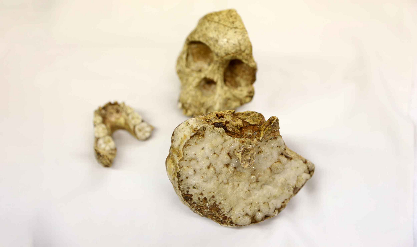
To a lot of anthropologists, the Taung endocast is one of their favorite fossils. It's a beautiful object in its own right. This endocast formed naturally when sediment entered the child's skull in its cave site and was hardened over time by minerals percolating into it. It preserves just over half the brain, most of the right side. On the side that was in direct contact with the skull, the reddish-brown, smooth surface of the endocast preserves the texture of the skull bones that encased it.
On the other side, where the sediment surface was exposed to air inside the skull, millimeter-sized crystals of calcite sparkle whenever light is shined across them. The skull had become a geode. Later most of the bone of the cranial vault was lost, either when the breccia was blasted or before.

The Taung endocast provides valuable information about the size and shape of the brain in this individual. Since it is the holotype of Australopithecus africanus it informs us about this species also. Raymond Dart, who described and first studied the Taung endocast, perceived several details as important in comparison with gorillas and chimpanzees. He emphasized that the endocast was larger than chimpanzees and nearly the size of a gorilla, much larger in body size. He observed that the Taung cerebral hemisphere was large in proportion to its cerebellum, and that the overall shape was “rounded and filled out”, which he interpreted as humanlike in contrast to the flatter shapes of great ape brains.
But endocasts are tough to interpret. The brain in humans and living great apes is not in direct contact with the inner surface of the skull, but is separated by thick membranes. Finding correspondences between bumps and grooves on the endocranial surface and the gyri and sulci of the brain is not always straightforward.
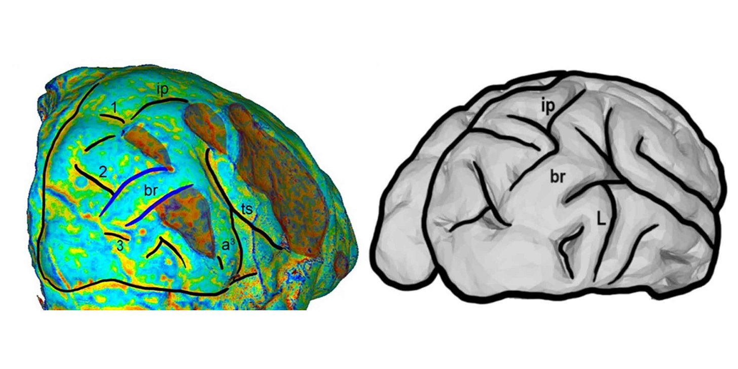
Over the years, one big area of disagreement emerged about how to interpret the Taung endocast. Dart identified a groove in the back part of the endocast as an anatomical feature known as the lunate sulcus. He saw this as significant as a marker of the extent of the parietal area where sensory input of different kinds is associated in the brain. Some later work disagreed with Dart's interpretation of the lunate sulcus, most notably the work of Dean Falk from the late 1970s and early 1980s.
Today the functional interpretation of this part of the brain differs somewhat from the 1925 view. The primary visual cortex in the occipital lobe of humans makes up a smaller fraction of the neocortex than in living great apes, and the parietal lobe of humans has expanded in relation to the primary visual cortex. In living chimpanzees and gorillas, a lunate sulcus often corresponds to the anterior boundary of the primary visual cortex. But in humans this is much more variable and a lunate sulcus often cannot be identified.

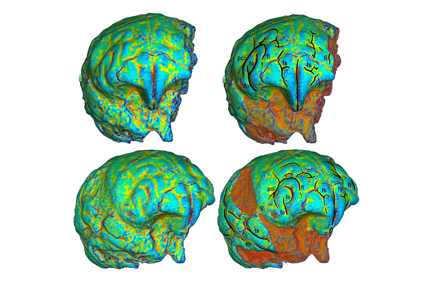
Shawn was able to bring to bear a large sample of chimpanzee brain scans, and a moderately large sample of human brain scans, to look at the variability of gyrus and sulcus position. The most interesting result of this work was a new interpretation of the lunate sulcus. Shawn saw that a “gyral bridge”, or a slight bulge interrupting the groove of a sulcus, is present in the majority of human brains where the lunate sulcus might otherwise be. This bridge connects occipital and parietal lobes and is also shared by the Taung endocast. It is present in a small fraction of chimpanzee brains but it is rare: only 1.8% of the hemispheres in the comparative dataset.
It's not evident whether this morphology of Taung shared with many humans has any correspondence to the functions of this part of the brain. It might be a side-effect of developmental processes, it might matter a great deal to connectivity. Since there are many living people who do not manifest this shape, it's hard to make any conclusions about the role of this morphology in our evolution, other than it became more common. But it does possibly provide a resolution for a long scientific dispute about how to interpret the shape evidence from the back part of the Taung endocast.
Overall, the new study shows that the Taung endocast shares some surface details with other great apes, while it shares some others with humans. This is not far off from Dart's assessment. But the curvature maps and broader comparative data in the paper should help other researchers use the data for replication, as well as making clear that the Taung specimen is situated within a tree of species, all of which exhibit some variability of brain form.


