Have Sahelanthropus and Orrorin been written out of existence?
A big argument about the so-called savanna theory comes with a surprising claim about the earliest possible hominin fossils.
Note: This post discusses current events and knowledge from the year 2014. Later discoveries and publications have added new information to the questions examined here, without changing the conclusions in this post. More recent posts discuss the Sahelanthropus postcranial remains described in 2022.
Barbara King devoted a recent NPR blog post to highlighting some professional acrimony in Current Anthropology: “Did Humans Evolve On The Savanna? The Debate Heats Up”.
In the Current Anthropology exchange, Manuel Domínguez-Rodrigo discusses the “savanna hypothesis” for the origin of hominin bipedal locomotion. The commentary of several experts follows the article, a regular feature of Current Anthropology. One of those experts is the noted paleoanthropologist Tim White, who argues strongly that the savanna hypothesis has been disproven by his work in the Middle Awash, on the habitat preferences of Ardipithecus.
In King’s view the resulting published exchange between White and Domínguez-Rodrigo crosses the line into unprofessional acrimony.
My view? Welcome to paleoanthropology.
I want to focus on a different aspect of the exchange. White’s comment on the paper includes the following passage:
Consequently, the simplistic narrative that hominid origins were initiated in open savannas created by climate change stands largely abandoned. Which available ecological habitat(s) among Africa’s diverse landscapes was favored by the earliest hominids (Ardipithecus subsumes Orrorin and Sahelanthropus; Haile-Selassie, Suwa, and White 2009)?
Wait a minute. This claim should be surprising to anyone following the literature on early hominins. Is it really viable to think that Ardipithecus should include both Orrorin and Sahelanthropus?
Origin of the claim
The claim originated in a paper by Yohannes Haile-Selassie, Gen Suwa and Tim White in 2004. There, they wrote about the variation in teeth attributed to Ardipithecus, noting that the hominin-like features of these teeth were shared with other fossil samples from the latest Miocene.
Metric and morphological variation within available small samples of late Miocene teeth attributed to A. kadabba, O. tugenensis, and S. tchadensis is no greater in degree than that seen within extant ape genera. Despite claims of molar enamel thickness differences among these late Miocene fossils, we question the interpretation that these taxa represent three separate genera or even lineages. Given the limited data currently available, it is possible that all of these remains represent specific or subspecific variation within a single genus.
That was the final paragraph of the paper, in some ways a shocker. Haile-Selassie, Suwa and White were claiming that the apparent diversity of early hominin fossil samples was mostly illusory. These different fossils, from Kenya, Chad and Ethiopia, all differed from Australopithecus and living apes in basically the same ways. Why not recognize that they are the same thing?
What they omitted from the paragraph is that some other samples of Miocene apes also differ from Australopithecus and living apes in similar ways. Primate paleontologists recognize the diversity of those lineages because they have other features that cannot be easily shoehorned into a single dental genus, and because many of them represent more of the skeleton than the teeth.
The weakness of the early hominin record (in 2004) was that except for the teeth, none of the available samples replicated the same parts. In 2004, the only parts of the Ardipithecus anatomy that had been described in any detail were its teeth. Orrorin had some teeth and a mandible along with three partial femora and a partial humerus. Sahelanthropus had a skull, with its teeth.
That's true of the published samples. From today's perspective, we know that much more material was available to some researchers but not to anybody else. Sahelanthropus had a partial femur and two ulnae; Ar. ramidus the partial “Ardi” skeleton along with other material. In 2004, nobody but the discoverers and their coworkers knew what these fossils might show, and they, including Haile-Selassie, Suwa and White, kept their cards close to their vests. So when these researchers asserted that the samples were consistent with variation in a single genus or species, independent scientists could evaluate this claim only on the basis of variation in the teeth.
The femur
That situation changed in 2009, as White and colleagues published a series of descriptions of the cranial and postcranial anatomy of Ardipithecus ramidus. Those descriptions are obviously relevant to the question of whether we can count these different samples as part of the same genus or species. Moreover, there has been a subsequent literature on some aspects of the anatomy of Ardipithecus, Sahelanthropus and Orrorin that offer additional points of comparison.
Lovejoy and coworkers (2009) described the femur of Ardipithecus, ARA-VP 1/701, emphasizing several morphological similarities with the Orrorin femora, in particular BAR-1002’00. The following paragraph gives a series of comparisons between the Ardipithecus femoral specimens and other early hominins, including BAR-1002’00:
In ARA-VP-1/701, the medial border of an obvious hypotrochanteric fossa homolog converges with the spiral line to form a markedly rugose, elevated plane on the posterior femoral surface, but their further course is lost to fracture. A similar morphology is visible on the ARA-VP-6/500 specimen. A broad linea of low relief is also clearly present in ASI-VP-5/154, assigned to Au. anamensis (34). Its morphology is reminiscent of that of A.L. 288-1, in which the linea is still notably broad, but contrasts with that of MAK-VP-1/1, which is more modern in form at 3.4 Ma (31). Because most of the length of the ASI-VP-5/154 shaft is preserved, its moderately elevated linea (~11.5 mm in breadth) is distinct and imparts a prismatic cross section at midshaft. Specimen BAR-1002'00 (Orrorin tugenensis) (35) presents obvious homologs to these structures. Moreover, both BAR-1002'00 and ASI-VP-5/154 exhibit an obvious homolog to the third trochanter, and neither shows any evidence of a lateral spiral pilaster.
The ARA-VP-1/701 and ARA-VP-6/500-5 femora from Aramis present few points of comparison because of the state of preservation. I cannot find the ARA-VP-6/500-5 femur illustrated anywhere except for the montage that shows the entire ARA-VP-6/500 skeleton. There is no photo of the femur fragment anywhere in the article by Lovejoy et al. 2009 or the supplementary information. Both pieces are described in the text as “proximal femur fragments” but neither preserves the proximal end; the better-illustrated ARA-VP-1/701 fragment ends just below the lesser trochanter.
The information from the Lukeino (Orrorin) femora is more complete: BAR-1002’00 includes a nearly complete proximal end and much of the shaft, and BAR-1003’00 replicates much of the anatomy of this specimen except for the head. These specimens show several features that link them with later hominins, including an elongated and anterio-posteriorly flattened femur neck, intertrochanteric line and obturator externus groove. The cortical bone distribution of the neck is
It is notable that the Aramis femoral fragments do not preserve the specific traits that most strongly link the Orrorin femur BAR-1002’00 to later hominins. As Lovejoy and colleagues (2009) discuss, there are similarities in the preserved portions between BAR-1002’00 and ARA-VP-1/701. The most striking of these is the “obvious homolog to the third trochanter” noted by Lovejoy and colleagues (2009). This is a marked, laterally projecting tuberosity for the attachment of the ascending tendon of the gluteus maximus muscle, on the lateral and posterior surface of the femur approximately at the same level as the lesser trochanter. Almécija and colleagues (2013) discuss this trait:
A different mode of bipedalism (from that of modern humans) practised by Orrorin (and Ar. ramidus) is consistent with the possession of a laterally protruding gluteal tuberosity below the greater trochanter (that is, small third trochanter, for insertion of the ascending tendon of the gluteus maximus) in combination with a ‘broad proto-linea aspera’. The latter arrangement indicates that a modern human configuration, consisting in a more posteromedial translation of the gluteus maximus insertion (functionally related to hypertrophy of the quadriceps at the expense of the hamstrings), had not yet occurred in the above-mentioned taxa.
The morphology of the gluteal tuberosity is shared by some early hominins but also with a number of Miocene apes. Pickford and colleagues (2002) emphasize that the form of this feature in BAR 1002’00 and BAR 1003’00 is different from Miocene apes except Ugandapithecus in being a relatively vertical crest, coalescing with the crest leading distally toward the linea aspera. They compare the form most closely with the Swartkrans femora.
Both Lovejoy and colleagues (2009) and Almécija and colleagues (2013) briefly discuss the “broad proto-linea aspera” morphology present in both the Aramis and Lukeino femora. This is the most hominin-like trait apparent in the Aramis femoral fragments, and the strongest argument that the femora reflect a functional or facultative bipedal ability in this primate. What weakens that argument for similarity is that we do not know the morphology of the common ancestors of hominins and chimpanzees. Almécija and colleagues (2013) note that the anatomical configuration represented by Miocene apes may have been closer to the pattern of the hominin-chimpanzee common ancestor than are living great apes:
Overall, our results agree with Napier’s viewpoint, according to which the morphological pattern of the proximal femur displayed by some Miocene apes is more likely to have been co-opted for bipedalism than the derived femoral morphology that is displayed by extant great apes. This does not necessarily imply a particularly close phylogenetic link between hominins and some of the Miocene apes included in our analyses. Rather, it would imply that Miocene apes would closely resemble the femoral morphotype of the last common ancestor between African apes and hominins (Fig. 5), whereas the morphology displayed by extant great apes would be much more derived—and probably homoplastic—as an adaptation for enhanced abduction during suspensory behaviours.
This is a similar argument to that proposed by Lovejoy and colleagues (2009): namely, that the anatomical pattern of living great apes is derived away from the hominin-chimpanzee common ancestor, and therefore those living apes are not good comparisons for early hominin locomotor anatomy. The implication of this argument is that we cannot assume that features shared by Lukeino and Aramis femoral fragments, but lacking in chimpanzees, are signs of common ancestry of these samples. They may reflect the anatomical inheritance of the common ancestors of hominins and other apes.
Kuperavage and colleagues (2010) considered the Orrorin femora in the context of the early hominin record, including Lovejoy and colleagues’ (2009) description of the Ardipithecus femoral anatomy:
At this stage in our knowledge of the first half of the human evolutionary record, approximately 6–3 Ma, there are more gaps than data points, and the data points that do exist mark populations that are at best known incompletely. The phrase “sparse and fragmentary” often is applied to this period, with complete justification. The situation is complicated still further by the non-comparable and otherwise unknown aspects of the fossils that have been discovered. Evidently much of a femoral diaphysis is known from deposits purportedly 7–9 Ma from Chad, but the specimen is still undescribed. The BAR 1002’00 and BAR 1003’00 femoral fragments comprise much of the proximal portion, with the region of the lesser trochanter well preserved. The next more recent sample, represented by ARA-VP-1/701, is not only separated by more than 1.5 Ma, but lacks nearly all of the lesser trochanter and more proximal portions. No os coxa accompanies BAR 1002’00 and 1003’00, while a heavily crushed specimen of this bone is represented by ARA-VP-6/500.
The absence of descriptions or publication of the Chad material makes it impossible to assess whether Sahelanthropus may conflict with the Ardipithecus or Orrorin femoral anatomy. I wrote about the purported Sahelanthropus femur in 2009: “Sahelanthropus: ‘The femur of Toumaï?’”
The skull
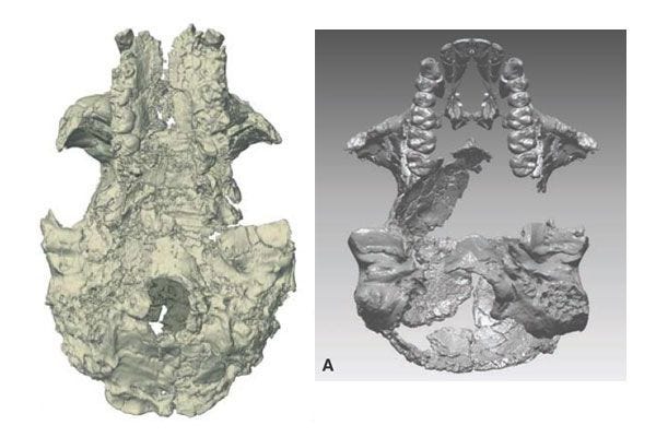
If we cannot compare the Sahelanthropus femur, we can certainly examine the cranium compared to the Aramis reconstruction. The Sahelanthropus and Ardipithecus crania look quite different from each other, even in the CT-based reconstructions. Here is TM 266-01-60-1 (abbreviated TM 266 as in Zollikofer et al. 2005), the skull of Sahelanthropus on the left (Zollikofer et al. 2005), compared to the composite reconstruction of the Ardipithecus cranium (Suwa et al. 2009) on the right. The two are approximately to scale. Suwa and colleagues (2009) give a series of linear measurements of the Ardipithecus cranial reconstruction, and provide equivalent estimates from the TM 266 reconstruction (in some cases estimated from photographs). I have aligned the two reconstructions approximately on the front of the foramen magnum. As is visually apparent, the TM 266 skull is substantially longer than the Aramis reconstruction. The two have approximately the same palate breadth, as estimated by Suwa and colleagues (2009).
Suwa and colleagues (2009) had to match the 3-dimensional configuration of the semicircular canals of the two temporal bones of the ARA-VP-1/500 specimen to find the relative position of these bones, and that information underlies the reconstruction of the cranial base here. The Aramis reconstruction has a slightly narrower cranial base than TM 266, but the TM 266 skull is overall much longer. Some of that greater length can be attributed to the extremely long nuchal plane of TM 266, which is extraordinary compared to later fossil hominins. Wolpoff and colleagues (including me) (2006) made the long nuchal plane one element of our argument that the Sahelanthropus skull morphology is outside the range of known hominins. That might appear to be a very important difference between the two crania. But in reality, the Ardipithecus cranial base and vault elements come from two different specimens, one of which (ARA-VP-1/500) was scaled to fit the other (ARA-VP-6/500). Neither specimen preserves the nuchal plane, which is a blank spot in the reconstruction.
If TM 266 should really be placed in Ardipithecus as suggested by White (2014), it is interesting to consider the Aramis cranial reconstruction with a nuchal plane oriented like TM 266. Below is a lateral view of both reconstructions, from Suwa et al. (2009) and Zollikofer et al. (2005):
It is not obvious from the visualization of the 3-d reconstruction how much of the curvature of the ARA-VP-6/500 parietals is actually present in the specimen, and how much of the appearance of curvature is the product of reconstruction. In lateral view, the other reason for the greater length of the TM 266 reconstruction is apparent: The skull has an extensive supraorbital torus which projects substantially anteriorly, which is very different from the Aramis reconstruction. By contrast, the Aramis reconstruction has a much more prognathic face, from the midface downward and particularly through the procumbent incisors. The profile of the TM 266 face is much more orthognathic. TM 266 is reconstructed with a broad, triangular nasal aperture with its superior border inferior to the orbits, and the Aramis reconstruction has a narrow nasal aperture with a rounded floor that extends superiorly above the base of the orbits.
There are, in other words, many visually striking differences between these reconstructions that reflect the basic architecture of the two skulls. We should keep in mind that they are reconstructions of crania that were highly distorted. The anatomical evidence makes it inevitable that TM 266 has a very long nuchal plane, for instance, and does not rule it out for the Aramis reconstruction. Both crania have been reconstructed with the biporion line approximately even with basion, and in both cases the interpretation of the reconstructions has favored a more vertical habitual posture.
It is not obvious to me that the two cranial reconstructions represent the same species or genus. At the same time, they are not obviously more different than two crania assigned to different species of Australopithecus, such as STW 505 and A.L. 444-2. It may be useful to think through the consequences of taking this assertion seriously. What would the Aramis reconstruction look like with a long, flat nuchal plane as in TM 266? What can we conclude about the anterior dentition of TM 266 if we use the Aramis dentition as a model?
Are Sahelanthropus and Orrorin sunk?
One way to tell whether this is a serious hypothesis is to examine the behavior of its authors. William Kimbel and colleagues, including Suwa and White (2014) recently published a study of the cranial base of Ardipithecus, in which they compared the ARA-VP-1/500 specimen with a number of other early hominins and living great apes. In this study, they somehow neglected to compare the specimen to the Sahelanthropus cranial specimen TM 266. Neither the word “Sahelanthropus” nor “Toros-Menalla” (the locality that produced the TM 266 skull) appears in the text.
This omission seems like a fairly obvious problem with that paper. Either the authors don’t really believe that the TM 266 skull belongs in Ardipithecus, or they believe it does belong in Ardipithecus and omitted half the basicranial sample. In either case, the TM 266 skull is obviously relevant; it is the only complete basicranium in the five million year span preceding the Aramis specimen. What should we conclude about the authors’ real opinion about Ardipithecus and Sahelanthropus?
I have worked with casts of the Orrorin material, and I have worked extensively with the published descriptions of the Sahelanthropus and Ardipithecus material. The Lukeino and Aramis femora are not identical, but the preserved portions of the Ardipithecus femoral specimens are not sufficient to demonstrate that they share the same derived features that apparently link Orrorin with later hominins. There cranial reconstructions from Toros-Menalla and Aramis are quite different from each other in both broad architectural features and in many details.
But only a handful of paleoanthropologists have access to accurate casts or three-dimensional CT-based models of the Ardipithecus and Sahelanthropus crania. Some of the relevant bone elements have never been illustrated in any journal, so it is impossible to verify some claims about them. The published descriptions notably omitted many standard details, such as standard dental measurements. We are in some ways no further along than we were in 2004 when Haile-Selassie, Suwa and White first published their claim that the dentitions of these samples represent a single species or genus. Some people know a lot about the anatomy of these samples, but they have not presented enough evidence for other scientists to evaluate their claims.
References:
Almécija S, Tallman M, Alba DM, Pina M, Moyà-Solà S, Jungers WL. 2013. The femur of Orrorin tugenensis exhibits morphometric affinities with both Miocene apes and later hominins. Nature Communications 4:2888. doi:10.1038/ncomms3888
Domínguez-Rodrigo, M. 2014. Is the “Savanna Hypothesis” a Dead Concept for Explaining the Emergence of the Earliest Hominins? Current Anthropology 55:59-81. doi:10.1086/674530
Guy F, Lieberman DE, Pilbeam D, Ponce de León, M, Likius A, Mackaye HT, Vignaud P, Zollikofer C, Brunet M. 2005. Morphological affinities of the Sahelanthropus tchadensis (Late Miocene hominid from Chad) cranium. Proceedings of the National Academy of Sciences of the United States of America, 102:18836-18841.
Haile-Selassie Y, Suwa G, White TD. 2004. Late Miocene teeth from Middle Awash, Ethiopia, and early hominid dental evolution. Science, 303(5663), 1503-1505. https://doi.org/10.1126/science.1092978
Lovejoy CO, Suwa G, Spurlock L, Asfaw B, White TD. 2009. The pelvis and femur of Ardipithecus ramidus: the emergence of upright walking. Science, 326(5949), 71-71e6. doi:10.1126/science.1175831
Kimbel WH, Suwa G, Asfaw B, Rak Y, White TD. 2014. Ardipithecus ramidus and the evolution of the human cranial base. Proceedings of the National Academy of Sciences, USA. 111:948–953. doi: 10.1073/pnas.1322639111
Pickford M, Senut B, Gommery D, Treil J. 2002. Bipedalism in Orrorin tugenensis revealed by its femora. Comptes Rendus Palevol 1:191-203. doi:10.1016/S1631-0683(02)00028-3
Suwa G, Asfaw B, Kono RT, Kubo D, Lovejoy CO, White TD. 2009. The Ardipithecus ramidus skull and its implications for hominid origins. Science 326:68-68e7. doi:10.1126/science.1175825
White T. 2014. Comment on "Is the 'savanna hypothesis' a dead concept for explaining the emergence of the earliest hominins?" Current Anthropology 55:75-76.
Wolpoff MH, Hawks J, Senut B, Pickford M, Ahern J. 2006. An ape or the ape: is the Toumaï cranium TM 266 a hominid? PaleoAnthropology 2006:36-50.
Zollikofer CP, Ponce de León MS, Lieberman DE, Guy F, Pilbeam D, Likius A, Mackaye HT, Vignaud P, Brunet M. 2005. Virtual cranial reconstruction of Sahelanthropus tchadensis. Nature 434:755-759. doi:10.1038/nature03397


