Tibia and fibula
A quick guide to the anatomy of the bones of the lower leg.
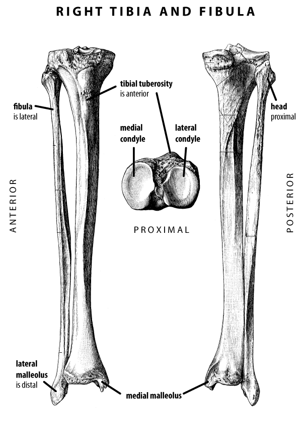
The tibia and fibula are the bones of the lower leg. The fibula forms the lateral part of the ankle joint, preventing dislocation in that direction.
The tibia is much larger and thicker than the fibula. On its proximal end, the tibia has two condyles that provide a platform on which the distal condyles of the femur sit during weight support. The anterior surface of the tibia lies just under the skin, and is often called the shin. At the proximal end of the anterior surface is the large tibial tuberosity, which most people can feel just below their kneecap.
The distal end of the tibia makes up much of the ankle joint, and the tibia has a distal projection on its medial side, called the medial malleolus, that can be felt on the inside surface of the ankle, and stabilizes the ankle in the medial direction.
The tibial tuberosity is on the front, or anterior aspect, of the tibia, and the medial malleolus is medial. These two features are good guides to determining whether a tibia is from the right or left side. Remember, it is the skeleton’s right or left, not yours as you look at the bone.