Assessing skeletal sex from the human pelvis
The pelvis is the most accurate indicator of sex in the human skeleton. Its central role in the birth process means that the pelvis has several shape differences between females and males. Learning these features is one of the fundamental bases of forensic identification.
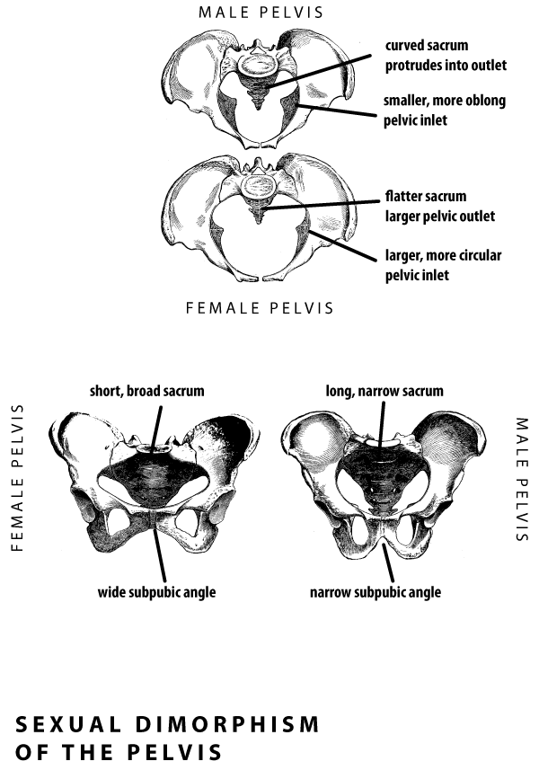
The lower part of the pelvis, called the true pelvis, contains the birth canal in females. The top of the true pelvis is defined by the pelvic inlet. The pelvic inlet is nearly circular in females, more oblong in males. The pelvis of males is overall larger, because the ilia (top part of the innominate bones) flare more extensively and the sacrum is longer.
The other features of the female pelvis tend to increase the size of the pelvic outlet, the space at the bottom through which the birth canal passes. Both pelvic bones and the sacrum have several shape differences in this area between males and females.
PubisThe anterior portions of the pelvic bones, called the pubes, comes together at the pubic symphysis. The inferior borders of the pubes form an angle, which is wider (around 90 degrees) in females, narrower (around 60 degrees) in males.Greater sciatic notchOn the posterior border of each innominate bone, the greater sciatic notch is wider in females, narrower in males.SacrumWider and shorter, with less curvature in females. Longer and more curved in males.
What to do: Examine the male and female pelves at this station. Use the other bones here to examine features related to sexual dimorphism. Try to identify male and female bones. Try seriating the bones to examine how the variation of different characteristics are related to each other.
Then go to one of the other tables, where pelvic bones have been arranged for you to determine sex. At that table, use the features of each bone to determine whether it is likely male or female. Write down the total number of male and female specimens in your determination, and leave those numbers with your TA.